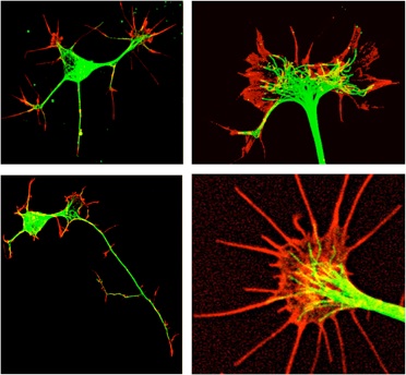
What is a growth cone? Normal sensation, motor control and behavior depend on correct wiring up of neuronal circuits during fetal development. The longest components of these circuits, the neuronal axons, sprout from immature neurons and elongate precisely through diverse tissues to reach synaptic partners located throughout the developing body. This embryonic axonal navigation is accomplished by the motile structure at the distal tip of an elongating neuronal axon, called a growth cone (Video 1). Like a bloodhound following a scent, a growth cone searches for and detects molecular signposts that are displayed throughout developing tissues. The growth cone responds to these signs by advancing, pausing and turning until it reaches its proper destination.
Growth cone history The Spanish neuroscientist Santiago Ramon y Cajal discovered growth cones in his anatomical studies of embryos, and in 1890 he published the first report and pictures of axonal growth cones (Figure 1). In 1907 at Yale University Ross Harrison observed the first living growth cones extended by neurons in tissue culture, and at the University of Virginia Carl Speidel first reported the activity of growth cones in vivo (Figure 2).
The pioneering work of these scientists inspires a legion of neuroscientists who investigate the basic mechanisms of growth cone behaviors, how growth cones navigate to their targets, and how growth cone behavior can be stimulated to promote axonal regeneration after injury or disease.
On the following pages you can read about how axons grow, how growth cones migrate, and how growth cone migration is regulated by guidance cues in developing tissues. Finally, growth cones and spinal cord regeneration are discussed.


